-
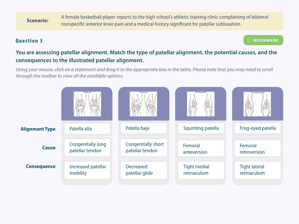
Congenitally short patellar tendon
-
Patella baja
-
Frog-eyed patella
-
Congenitally long patellar tendon
-
Tight lateral retinaculum
-
Femoral anteversion
-
Tight medial retinaculum
-
Femoral retroversion
-
Patella alta
-
Increased patellar mobility
-
Squinting patella
-
Decreased patellar glide
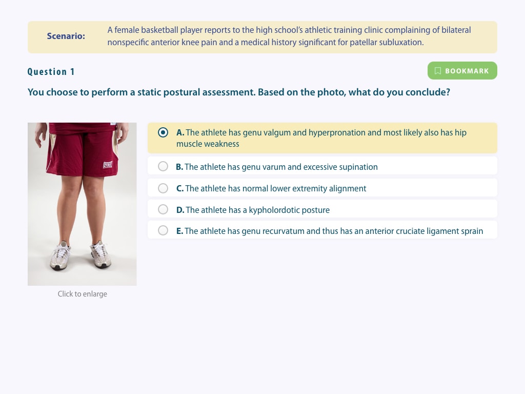
Scenario:
A female basketball player reports to the high school’s athletic training clinic complaining of bilateral nonspecific anterior knee pain and a medical history significant for patellar subluxation.
Question 1
You choose to perform a static postural assessment. Based on the photo, what do you conclude?

Click to enlarge
The athlete has genu valgum and hyperpronation and most likely also has hip muscle weakness
The athlete has genu varum and excessive supination
The athlete has normal lower extremity alignment
The athlete has a kypholordotic posture
The athlete has genu recurvatum and thus has an anterior cruciate ligament sprain
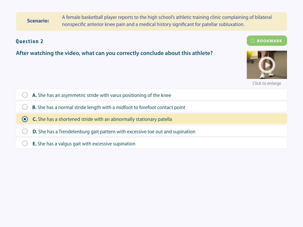
Scenario:
A female basketball player reports to the high school’s athletic training clinic complaining of bilateral nonspecific anterior knee pain and a medical history significant for patellar subluxation.
Question 2
After watching the video, what can you correctly conclude about this athlete?

Click to enlarge
She has an asymmetric stride with varus positioning of the knee
She has a normal stride length with a midfoot to forefoot contact point
She has a shortened stride with an abnormally stationary patella
She has a Trendelenburg gait pattern with excessive toe out and supination
She has a valgus gait with excessive supination
Scenario:
A female basketball player reports to the high school’s athletic training clinic complaining of bilateral nonspecific anterior knee pain and a medical history significant for patellar subluxation.
Question 3
You are assessing patellar alignment. Match the type of patellar alignment, the potential causes, and the consequences to the illustrated patellar alignment.
Using your mouse, click on a statement and drag it to the appropriate box in the table. Please note that you may need to scroll through the toolbar to view all the available options.

|

|

|

|
|
|---|---|---|---|---|
|
Alignment Type |
|
|
|
|
|
Cause |
|
|
|
|
|
Consequence |
|
|
|
|
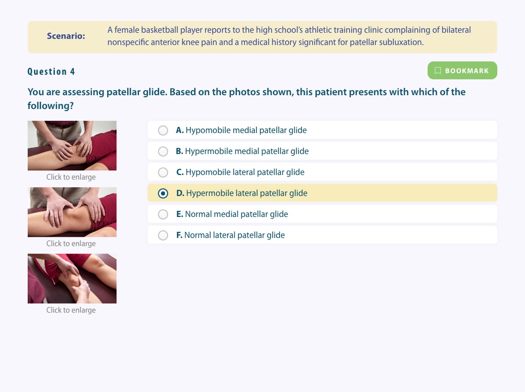
Scenario:
A female basketball player reports to the high school’s athletic training clinic complaining of bilateral nonspecific anterior knee pain and a medical history significant for patellar subluxation.
Question 4
You are assessing patellar glide. Based on the photos shown, this patient presents with which of the following?

Click to enlarge
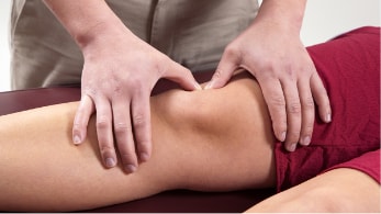
Click to enlarge
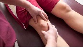
Click to enlarge
Hypomobile medial patellar glide
Hypermobile medial patellar glide
Hypomobile lateral patellar glide
Hypermobile lateral patellar glide
Normal medial patellar glide
Normal lateral patellar glide
Scenario:
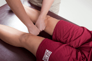
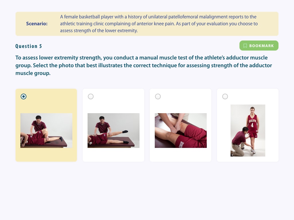
A female basketball player with a history of unilateral patellofemoral malalignment reports to the athletic training clinic complaining of anterior knee pain. As part of your evaluation you choose to assess strength of the lower extremity.
Question 5
To assess lower extremity strength, you conduct a manual muscle test of the athlete’s adductor muscle group. Select the photo that best illustrates the correct technique for assessing strength of the adductor muscle group.


Scenario:
A female basketball player with a history of unilateral patellofemoral malalignment reports to the athletic training clinic complaining of anterior knee pain. As part of your evaluation you choose to assess strength of the lower extremity.
Question 6
To assess lower extremity strength, you conduct a manual muscle test of the athlete’s semimembranosus and semitendinosus. The athlete’s lower leg must first be correctly positioned. From the choices below select the arrow that, when placed on the metatarsal heads, best represents the direction you will rotate the lower leg to achieve correct test position for this manual muscle test.
First, use your mouse and select an arrow from the toolbar. Only one arrow can be selected. Second, click the metatarsal heads in the image to indicate the correct direction of rotation. You may reposition the circle until you are satisfied with its placement. When you have completed your assessment, click the SUBMIT RESPONSE button to submit the question for scoring.


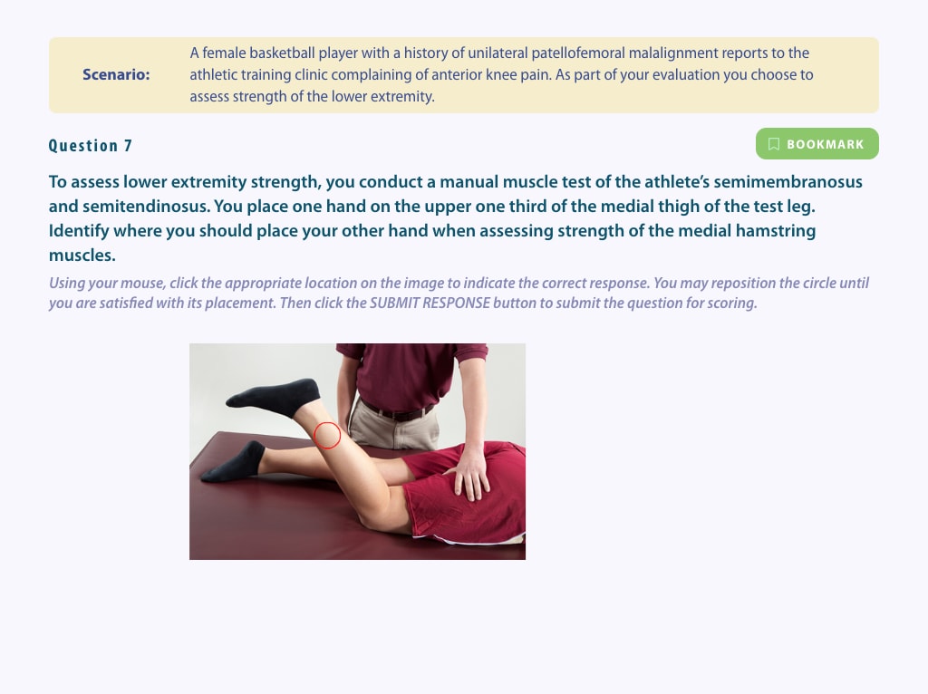
Scenario:
A female basketball player with a history of unilateral patellofemoral malalignment reports to the athletic training clinic complaining of anterior knee pain. As part of your evaluation you choose to assess strength of the lower extremity.
Question 7
To assess lower extremity strength, you conduct a manual muscle test of the athlete's semimembranosus and semitendinosus. You place one hand on the upper one third of the medial thigh of the test leg. Identify where you should place your other hand when assessing strength of the medial hamstring muscles.
Using your mouse, click the appropriate location on the image to indicate the correct response. You may reposition the circle until you are satisfied with its placement. Then click the SUBMIT RESPONSE button to submit the question for scoring.

|
Question Number |
Your Score |
Possible Score |
Answer Key |
|---|---|---|---|
|
1 |
0 |
1 |
|
|
2 |
0 |
1 |
|
|
3 |
0 |
12 |
|
|
4 |
0 |
1 |
|
|
5 |
0 |
1 |
|
|
6 |
0 |
2 |
|
|
7 |
0 |
1 |
|
|
Total Points |
0 |
19 |
|
|
Percentage |
|
||